The Electron Microscopy and Microanalysis Laboratory is a Joint Research Laboratory CNR-IGG / Department of Earth Sciences, University of Turin for the observation and detailed analysis of the morphology and chemical composition of solid materials.
The Laboratory
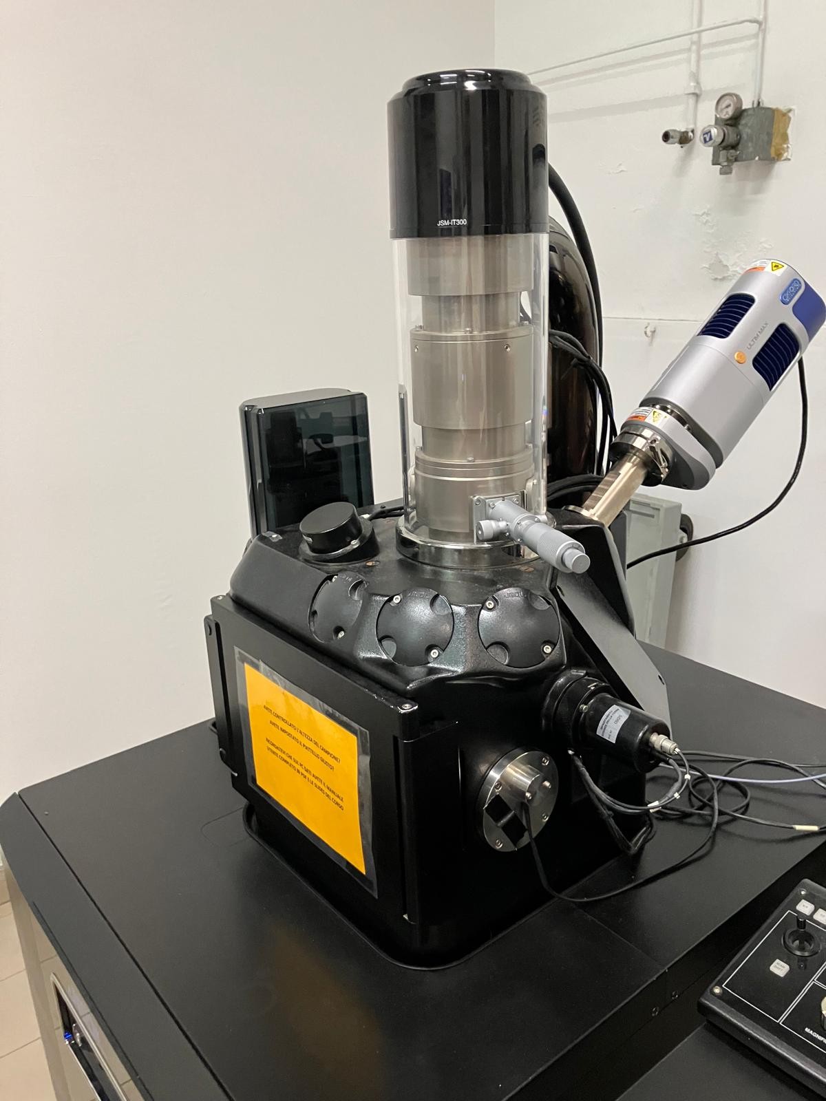
The laboratory uses the JEOL JSM IT300LV (High Vacuum – Low Vacuum) Scanning Electron Microscope equipped with an OXFORD Aztec Energy SDD ULTIMAX65 EDS microanalysis unit.
The scanning electron microscope (SEM) uses an electron beam to generate high-resolution images of the sample surface, allowing microscopic structures to be observed with great depth of field.
The EDS (Energy Dispersive Spectroscopy) detector enables elemental microanalysis: it detects the X-rays emitted by the sample under the electron beam and identifies the chemical elements present and their distribution.
Aztec Energy software allows for image acquisition and processing and qualitative and quantitative processing of spectra from point, line, or area analyses.

The instrumental apparatus is equipped with:
- electron gun for producing and focusing the electron beam
- a chamber for housing the sample
- a vacuum production system
- micrometric movement systems,
- rivelatori elettronici e a raggi X,
- computers with the appropriate Aztec software for the acquisition and processing of the acquired data.
This type of laboratory is essential for materials characterization in the fields of planetary science research, engineering, biomedicine, and materials science.
Instruments
The equipment available to the laboratory is:
- SEM Electronic Microscope – JEOL IT-300LV, (High Vacuum – Low Vacuum 10/650 Pa – 0.3-30kV);
- Microanalysis using the OXFORD Aztec Energy Energy Dispersive Spectrometer equipped with:
- OXFORD ULTIMAX65 SDD type detector, with active area of 65 mm2 and energy resolution of at least 127 eV (on Mn Kalpha);
- Aztec Live software for live analysis, EDS mapping, quantitative or standard line-scan point analysis less
- On-line and off-line processors for punctual, linear and areal microanalytical reprocessing;
- The metalizing and graphitizing equipment present in the laboratory includes:
Staff and Contacts
The laboratory staff is composed of:
- Dr. Gloria Vaggelli (Scientific Director for CNR-IGG)
- Dr. Emanuele Costa (Scientific Director for DST-UNITO)
Contacts:
- Dr. Gloria Vaggelli: +39 011 6705119, Gloria.vaggelli@cnr.it
Scientific Projects and Interests
The Laboratory aims to support multidisciplinary research activities, with particular attention to:
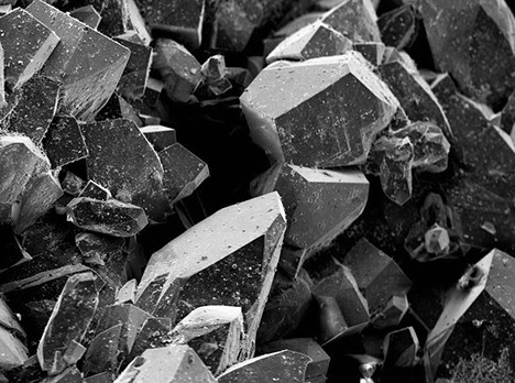
- Studies on minerals, rocks, and synthetic analogues, for compositional, morphological, and morphometric characterization for petrological purposes. (Fig. 1)
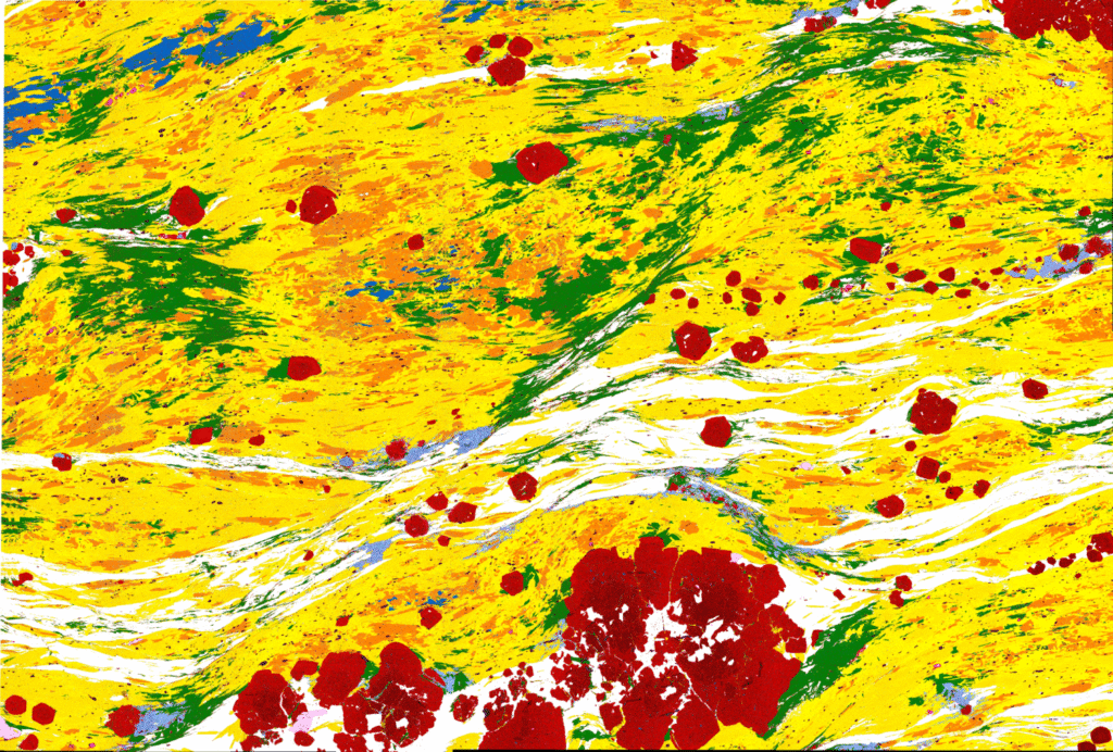
- Methodological studies for the development of techniques that use compositional maps and imaging analyses of minerals and rocks. (Fig. 2)
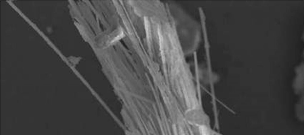
- Characterization of asbestos, asbestiform minerals, and silica dust present in airborne particulate matter and biological tissues. (Fig. 3)
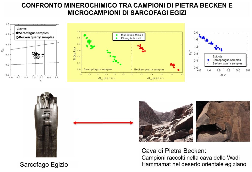
- Minerochemical characterization of ceramics, glass, coins, and artifacts of historical-archaeological interest. (Fig. 4)
The scientific activities referred to in the points above are expected to fall within the general macro-themes inherent to planetary systems, and with special reference to the Earth, and include issues relating to the environment, health and cultural heritage.
Publications
- Cossio R., Ghignone S., Borghi A., Corno A., Vaggelli G. (2024). A supervised machine learning procedure for EPMA classification and plotting of mineral groups. Applied Computing and Geosciences 23 (2024) 100186. https://doi.org/10.1016/j.acags.2024.100186
- Ghignone, S., Prencipe, M., Bruno, M., Boero, F.,Costa, E., Scaramuzzo, E. (2023). The Raman spectrum of florencite-(REE) [REEAl3(PO4)2(OH)6]: An integrated experimental and computational approach. Journal of Raman Spectroscopy, 55 (3) 394-405. https://dx.doi.org/10.1002/jrs.6640.
- Visonà, S.D., Capella, S., Borrelli, P., Villani, S., Favaron, Kurzhunbaeva, Z., Colosio, C., Belluso, E. (2023). Asbestos burden in lungs of non-occupationally exposed women from Broni (Pavia, Italy): a postmortem SEM-EDS study. Journal of Thoracic Disease 15(12):6555-6569. https://dx.doi.org/10.21037/jtd-23-1061.
- Gambino, F., Glarey, A., Cossio, R., Appolonia, L., d’Atri, A., Borghi A. (2022). SEM-EDS characterization of historic mortar as a tool in archaeometric study: an updated analytical protocol tested on the Roman theatre of Aosta (NW Italy). Archaeological and Anthropological Sciences, 14, 179. https://doi.org/10.1007/s12520-022-01645-9.
- Vaggelli, G., Es Sebar, L., Borghi, A., Cossio, R., Re, A., Fantino F., Lo Giudice, A. (2019). Improvements to the analytical protocol of lapis lazuli provenance: First study on Myanmar rock samples. Eur.Phys.J.Plus(2019) 134:(104) 1-15 https://doi.org/10.1140/epjp/i2019-12523-4.
- Costa, E., Bittarello, E., Navone, R. (2017) Chalcedony beads coated with titanium nitride. In GEMS & GEMOLOGY 53 (2) (Summer 2017).
- Gulmini M., Roselli G., Scognamiglio F., Vaggelli G. (2015). Composition and microstructure of maiolica from the museum of ceramics in Ascoli Piceno (Italy): evidences by electron microscopy and microanalysis. Applied Physics, 120 (4), p. 1643-1652; doi 10.1007/s00339-015-9376-9.
- Serra M., Borghi A., D’Amicone E., L. Fiora, Mashali O., Vigna L. and Vaggelli G (2010): Black and red granites in the Egyptian antiquity museum of Turin, a minero-petrographic and provenance study, Archaeometry VOL 52 (6): 962-986. DOI: 10.1111/j.1475-4754.2010.00535.x.