
The Laboratory of Microanalysis for Electron Microprobe (LaMa) is a joint laboratory of IGG-CNR / Department of Earth Sciences, University of Florence
The Laboratory
Electron microprobe analyses allow for the in situ chemical composition of any solid, inorganic material, whether crystalline or amorphous, natural or artificial. This instrument has many applications, ranging from topics closely related to Earth Sciences research to archaeometry and cultural heritage, and even materials science and engineering.
The LaMa microanalysis laboratory is a laboratory shared with the Department of Earth Sciences, where the main instrumentation was strengthened in 2020 with the purchase of the new JEOL JXA-8230 electronic microprobe.
The electron microprobe is the primary tool for obtaining the in situ chemical composition of any solid, inorganic material, whether crystalline or amorphous, natural or artificial, with high spatial resolution (in the micron range). The instrument is specifically designed for the detailed quantitative analysis of all elements with Z>3 (essentially from Be to U) with a broad analytical capacity that allows for the detection of major (>1 wt%), minor (0.1-1 wt%) and, in some appropriately managed cases, trace (<0.1 wt%) contents, after calibration with specific certified international standards.
The laboratory's activity focuses primarily on the analysis of silicate minerals, oxides, sulfides, phosphates, metals, and carbonates present in terrestrial and extraterrestrial rocks with the aim of obtaining information on the geological processes that generated them.
Analyses of specific matrices have also been implemented for applications in the fields of archaeometry (artificial glasses, terracottas, ceramics), paleontology (bones, teeth), and crystal chemistry, as well as microanalysis of industrial materials (e.g., cements, alloys) for studies related to Materials Sciences.

Instruments
The laboratory is equipped with:
- JEOL JXA-8230 Electronic Microprobe.
The instrument, installed in 2020, is equipped with five wavelength-dispersive spectrometers (WDS), each housing several analyzer crystals, some of which are dedicated to the analysis of light elements (1: TAPJ, LDE1, PETJ, LDEB; 2: LIFL, PETL; 3: LIFH, PETH; 4: LIF, PETJ; 5: TAPJ, LDE2), and detectors consisting of sealed gas-flow proportional counters. A solid-state energy-dispersive detector (EDS) allows instantaneous acquisition of the emission spectrum and rapid identification of the elements present in the sample.
The electron source can be a W filament (ideal for compositional analyses) or a LaB6 crystal (potentially useful for high-resolution mapping applications, as it produces a particularly thin beam shape, increasing spatial resolution).
In its standard configuration, the instrument currently works with a W filament.
An additional ion pump allows for high vacuum conditions in the source, which guarantee greater stability of the incident electron beam and consequently higher analytical quality.
The analytical potential of this new tool therefore allows us to:
- analyze light elements starting from Be, thanks to the presence of specific and dedicated analyzer crystals (LDE1; LDEB; LDE2);
- have reduced analytical times, thanks to the presence of 5 WDS spectrometers;
- have greater sensitivity that allows the detection of trace elements in the order of hundreds of ppm, thanks to the presence of spectrometers with specific geometries;
- perform high spatial resolution WDS compositional maps thanks also to the possibility of inserting a LaB6 source;
- use an EDS system with a high-performance detector that allows obtaining a complete compositional spectrum in a short time, potentially resolute even at low energies (e.g. elements such as O, F, B), and indicative maps of the elemental distribution in the different samples;
The sample is viewed through a digital SEM screen, both in backscattered electrons (BSE), obtaining a compositional image in grayscale proportional to the electron density (mean atomic weight) of the material, and in secondary electrons (SE), obtaining a morphological image of the sample's surface. A camera connected to another monitor allows the sample to be focused and observed in reflected and/or transmitted light, thanks to an additional lens (OMT) that can be inserted into the chamber beneath the sample holder (only for samples mounted on petrographic sections).
The integrated software allows you to program specific analytical routines for the different phases being investigated, customizing them to meet your needs (e.g., element order, counting times, current intensity, etc.). It also allows you to program serial sequences that can be run automatically, storing multiple analysis points. This optimizes machine time and increases the number of analyses that can be performed.
- Leica EM ACE 200 Metallizer
- JEOL JEE-4X Graphite Metallizer
- Zeiss Jenalab optical microscope in reflected and transmitted light
Staff and Contacts
Dr. Eleonora Braschi (CNR Researcher – Supervisor)
Dr. Andrea Orlando (CNR Researcher)
Prof. Simone Tommasini (PA UniFi associated with IGG – Manager)
Phones
+39 055 2757565 (Dr. Eleonora Braschi)
+39 055 2757510 (Dr. Andrea Orlando)
+39 055 2757793 (Laboratory)
E-mail:
Methods and Applications
Given the instrument's broad analytical capacity and the detail that this analysis method allows, the possible applications are very diverse, as are the analytical protocols developed for the different types of matrix or elements to be analyzed.
The analytical conditions generally used are 15kV acceleration, 20nA current for anhydrous silicates, 10nA for natural glasses, and 5nA for hydrated silicates, with beam dimensions varying from 3µm to 5µm and 10mm depending on the matrices analyzed. Higher acceleration voltages (20kV) and variable currents are used for more specific phases such as sulfides or phosphates rich in rare earth elements (e.g., monazites).
Counting times vary depending on the elemental abundances to be analyzed and the needs; generally, 15 s of counting are set on the peak and 7 s on each background for major elements, with the exception of Na (10 s on the peak and 5 s on each background) to limit diffusion effects. Longer counting times, from 30 s to 40 s on the peak and 15-20 s on each background, are used for minor elements.
The order of analysis and the distribution of the elements in the different spectrometers is specific and customized for the different phases/matrices analyzed and involves 2 to 3 counting cycles for each analysis. In silicates, Si, Na, and K are always analyzed first to minimize alkali diffusion effects and the resulting matrix effect on the silicon.
The correction method applied for the matrix effect is of the ZAF type for silicates and oxides or PAP for sulphides, carbonates and phosphates.
The electron microprobe analytical method allows for in situ, or point-by-point, analysis. It does not allow for bulk analysis of a sample, but provides detailed compositional information, allowing for the exploration of small-scale phase composition variability.
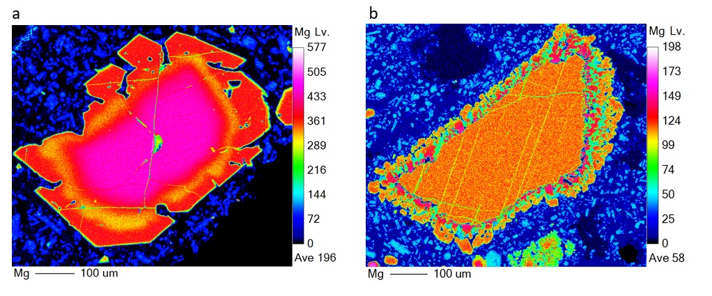
Alternatively, the instrument can provide semi-quantitative, high-resolution compositional maps over variable areas of the test sample for a selection of elements.
The main applications on which the laboratory's activity is mainly concentrated:
- Applications to Earth Sciences, particularly to the mineralogy and petrology of both igneous and metamorphic rocks, and, secondarily, sedimentary rocks, through the analysis of mineral matrices (mainly silicates, but also oxides, sulfides, carbonates, and phosphates) and glasses present in terrestrial and extraterrestrial rocks (lunar and Martian meteorites). The objectives are primarily related to obtaining information for the investigation of the geological processes that generated them, the differentiation processes, and the characteristics of the systems in which these processes occur, as well as the timescales over which these processes developed, providing the basis for more advanced applications (e.g., U-Th dating of monazites, zircons, titanites; diffusion times);
- applications in the archaeometric field, with analysis of ancient glass in order to identify the constituent components (raw materials) and their origin, metal alloys, terracotta and ceramics;
- application in the field of paleontology through the analysis of mineralized portions of bones or teeth, capable of providing information on the life and habits of animals that lived in the past, or shells of marine organisms with applications for paleoecology;
- applications in the field of crystal chemistry, as well as new materials with analysis of specific matrices, for application in the most modern fields of new technologies and green energy.
To meet the various analytical needs, the laboratory has equipped itself with some specific sets of certified standards:
- Astimex MIN standards used for the calibration of the elements analyzed in silicates and oxides and secondarily in sulfides, phosphates and carbonates;
- Astimex MET standards used for the calibration of analyzed elements in metal alloys;
- P&H standards for the calibration of elements analyzed in sulfides;
- P&H Standards for Rare Earth Calibration;
- International standards (Smithsonian, BioRad) and others to test the quality of instrument calibration and the reproducibility of analyses over time.
For correct analysis, the sample requires specific preparation. The samples must be well-polished in the form of thin sections measuring 48x28mm, or as 1-inch (25.4mm) embedded specimens, with a variable thickness, but preferably 9 or 5mm. Each sample is subjected to metallization by spraying a thin layer of graphite (approximately 28nm).


Scientific Projects and Interests
The laboratory has been actively used in several scientific projects, including:
- PRIN 2017 project (MIUR: 2017P9AT72 PE10)
- PRIN 2017 project (MIUR: 20178LPCPW PE10)
- International Exchanges 2023 Cost Share project Royal Society UK (IECÄR2Ä232044)
- Nanosolid project (Tuscany Region partnership)
- Space It Up project (ESA partnership)
The laboratory is attended by researchers and professors from various Italian and foreign research institutions and universities, upon reservation.
The laboratory's primary scientific interests are in the petrology of igneous rocks, studying the genesis and evolution of magmas and volcanic systems through the careful analysis of mineral phases and glasses. This is complemented by the characterization of extraterrestrial rocks through detailed investigations of specific phases such as sulfides, phosphates and phosphides, oxides, and silicates that distinguish them.
Further interests are focused on the analysis of rare earth phases such as monazites, zircons, titanites for thermochronological applications.
Publications
- Braschi et al., 2025. Eur. J. Mineral., 37, 793–817. https://doi.org/10.5194/ejm-37-793-2025.
- Corvò et al., 2023. Earth and Planetary Science Letters, 620, 118349.https://doi.org/10.1016/j.epsl.2023.118349.
- Orlando et al., 2025. Geoheritage 17:179, doi.org/10.1007/s12371-025-01233-y.
- Gliozzo et al., 2023. Microchemical Journal 189, 108511. https://doi.org/10.1016/j.microc.2023.108511.
- Gliozzo et al., 2024. Archaeological and Anthropological Sciences, 16:114. https://doi.org/10.1007/s12520-024-02017-1.
- Gliozzo et al., 2025. Archaeological and Anthropological Sciences, 17:207. https://doi.org/10.1007/s12520-025-02317-0.
- Gualtieri et al., 2024. Eur. J. Mineral., 36, 749–765. https://doi.org/10.5194/ejm-36-749-2024.
- Langone et al., 2025. Geological Magazine 162(e18): 1–24. https://doi.org/10.1017/S0016756825100022.
- Orlando et al., 2025. International Journal of Earth Sciences. https://doi.org/10.1007/s00531-025-02493-3.
- Pisello et al., 2023. Minerals, 13, 170. https://doi.org/10.3390/min13020170.
- Tursi et al., 2020. J Metamorph Geol., 38:451–469. DOI: 10.1111/jmg.12528.
- Tortelli et. al., 2024. Contributions to Mineralogy and Petrology, 179:87. https://doi.org/10.1007/s00410-024-02164-7
- Vettori et al., 2025. Journal of Archaeological Science: Reports 67: 105396. https://doi.org/10.1016/j.jasrep.2025.105396